Our Research

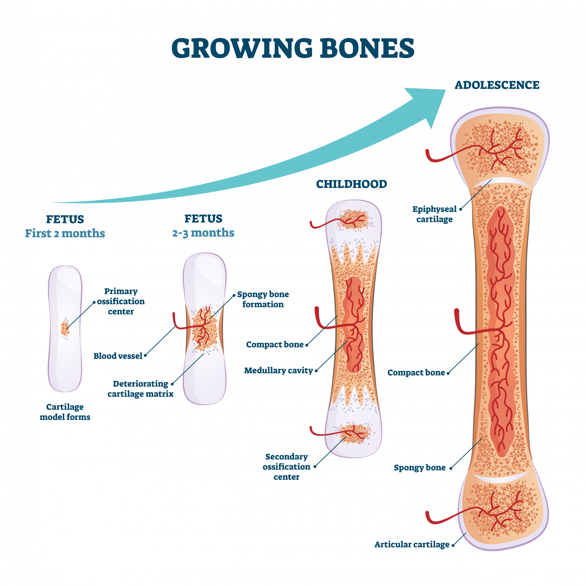
Throughout fetal development, an early cartilage template prefigures the shape of the future bones and provides progenitors for skeletal growth. Osteogenesis sprouting in the center of the skeletal elements, gradually remodels the cartilaginous anlagen and reduces it to a smaller structure, located near the ends of the skeletal elements. A secondary ossification center (SOC) further divides the remaining cartilage into a narrow band, called the growth plate (GP), located deeper into the bone, and another band, the articular cartilage (AC), located on the surface of the skeletal elements. The growth plate cartilage supports the growth and elongation of the limbs until puberty, when its closure halts any further skeletal growth. The articular cartilage provides elasticity and resistance to compressive loads and allows smooth, pain-free joint movements. Both GP cartilage and articular cartilage have a similar architecture, consisting of layers of immature and hypertrophic (collagen 10+) chondrocytes.
Current areas of research include:
Stem cell guided cartilage repair after injury
Children who have sustained injuries to the GP often experience growth arrest and may suffer from significant disability in adulthood due to malalignment of the appendicular skeleton, leg length inequality, and articular joint dysplasia. This highlights the need for novel therapeutic strategies to repair the tissue at the injury site and to allow the GP cartilage to resume growth. We seek to develop new strategies for stem cell guided cartilage repair after injury.
Understanding Osteoarthritis progression in the synovial joints
Osteoarthritis (OA) is a painful joint disease, characterized by progressive and irreversible deterioration of the cartilage. There is no cure for OA. Current treatments provide, at best, relief from pain and inflammation associated with the more advanced phases of disease but they do not target the OA progression because the molecular mechanisms are not well understood. We want to elucidate the sequence of events that leads to joint cartilage damage in hopes of designing new therapies for tissue repair.
Transcriptional control of skeletal development
In endochondral ossification, which takes place in the axial and appendicular skeleton, bone formation occurs indirectly via formation of a transient cartilage template. The mesenchymal progenitor cells differentiate into chondrocytes, and lay down a cartilage anlagen, which is subsequently replaced by bone. Using a combination of genomics, genetics and biochemistry, we want to study the critical interactions between various transcription factors and determine the gene signature of the cartilage/bone interface to understand how skeletal elements grow.